Pyeloplasty
Dr. Shaw is proud to be one of a few Urologists in Central Texas to offer expertise in da Vinci (robotic) repair of obstructed, or blocked kidneys. Where this was once a major procedure with an incision from the front to back under the ribs, we replace this with 4 tiny incisions the width of your thumb.

Pyeloplasty Information
- What happens under normal conditions?
- What is ureteropelvic junction (UPJ) obstruction?
- What are the symptoms of ureteropelvic junction (UPJ) obstruction?
- How is ureteropelvic junction (UPJ) obstruction diagnosed?
- How is ureteropelvic junction (UPJ) obstruction treated?
- What can be expected after treatment for ureteropelvic junction (UPJ) obstruction?
- Pre-Procedure
- Procedure
- Post Procedure
- Advice After da Vinci Robotic Pyeloplasty
- Follow-up
- Long Term
- Videos
What happens under normal conditions?
Kidneys produce urine by filtering the blood and removing wastes, salts and water. The urine must then drain from the kidney through an internal collecting system that ends in a funnel-shaped structure called the renal pelvis and into a natural tube called the ureter. Each kidney must have one functional ureter to carry the urine from the kidney to the bladder.
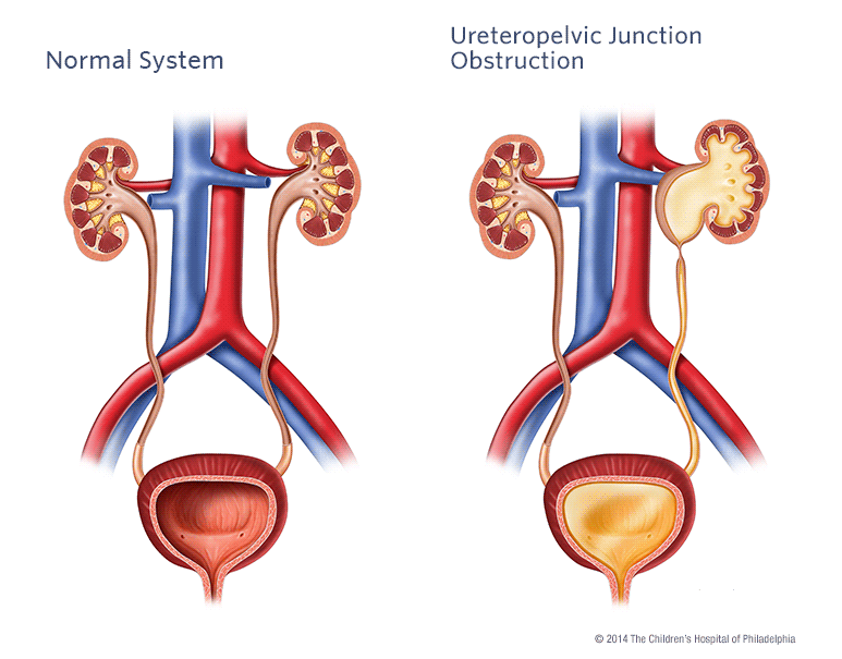
What is ureteropelvic junction (UPJ) obstruction?
The most common cause of obstruction (blockage) in the urinary tract is a congenital obstruction at the point where the ureter joins the renal pelvis — the ureteropelvic junction (UPJ). This problem occurs in approximately one in 1,500 children. These obstructions develop prenatally as the kidney is forming and today most are diagnosed on prenatal ultrasound screening. In UPJ obstruction, the kidney produces urine at a rate that exceeds the amount of urine able to drain out of the renal pelvis into the ureter and this causes accumulation of urine within the kidney. This accumulation, also called hydronephrosis, is easily visible on ultrasound and often allows the physician to predict the presence of UPJ obstruction before the baby is born. Although encountered less frequently in adults, UPJ obstruction may occur as a result of kidney stones, previous.
What are the symptoms of ureteropelvic junction (UPJ) obstruction?
Since the development of ultrasound and its widespread use to screen the unborn child, most UPJ obstructions are identified long before birth. After birth, symptoms of UPJ obstruction may be an abdominal mass, a urinary tract infection with fever, flank pain especially with increased fluid intake, stones and bloody urine. Patients with UPJ obstruction may also have pain without an infection. Some UPJ obstructions are irregular in nature and urine may drain normally at one time and be completely obstructed at others producing sporadic pain.
How is ureteropelvic junction (UPJ) obstruction diagnosed?
While ultrasound is a very useful screening test it is not diagnostic of UPJ obstruction. In order to make the diagnosis it is necessary to perform a functional test or one that measures the ability of the kidney to produce and drain urine. The classic examination is called the intravenous pyelogram (IVP), or a CT with IV contrast. In this test, a dye is injected into the blood stream and the kidneys remove this substance from the blood. The dye passes into the urine and eventually out of the bladder. The dye is visible on X-ray and the physician can see the shape of the kidney, renal pelvis and ureter. Another useful examination is the furosemide renal scan. This test is done in a similar fashion to the excretory urogram except that a radioactive material is used instead of X-ray dye. The material can be followed with a special camera and this test can give more accurate information about kidney function and drainage.
How is ureteropelvic junction (UPJ) obstruction treated?
Maximum Results, Minimal Incisions.
Dr. Shaw is proud to offer the latest technology for management of urologic disorders with robotics. Dr. Shaw was amongst the first surgeons in the area to offer this option to patients, and began his training in robotics in 2000 while in residency at Tulane, the first robotics program in the Gulf Coast region. Dr. Shaw has been chosen since 2004 to be one of a select group of surgeons from Intuitive Surgical, to be a nationwide proctor of da Vinci Robotic Surgery. Dr. Shaw routinely participates in national and international events for robotic surgery, keeping with the latest technology in this rapidly expanding area of Urology. The classic treatment of UPJ obstruction is an open operation to remove the UPJ and to reattach the ureter to the pelvis of the kidney creating a wide junction between the two. This operation, called a pyeloplasty, allows rapid and easy drainage of urine produced by the kidney and relieves symptoms and the risk of infection. The procedure usually has a success rate in excess of 95 percent with one operation. Hospitalization after surgery depends on age of the patient. Newer treatment of UPJ obstruction involves minimally invasive surgery. Dr. Shaw is able to replicate the same procedure that is done with a large (open) incision with the latest technology. Dr. Shaw is proud to offer da Vinci laparoscopic, robotic repair of UPJ obstruction. What is da Vinci Robotic Surgery? Simply put, the reason why we used to make large incisions for major surgeries was so we could put our hands inside the body. With the the advent of robotics, we perform the same surgery, arguably better, with smaller incisions, because instead of the surgeon’s hands, we only need to make a few small 1/2 inch incisions to insert the robotic hands. From there, we use 3-D vision, High Definition (HD) Cameras and the latest technology to translate the surgeons hand movements to the robotics. Think Wii for robotic surgeons! All this technology translates to more precise movements, vision that is magnified in 3-D and other benefits. Results? My patients rarely require blood transfusions, have less pain and smaller incisions than open surgery, and return home in 24-48 hours.
What can be expected after treatment for ureteropelvic junction (UPJ) obstruction?
Patients usually recover quickly from any of the procedures but some have pain for a few days following surgery and occasionally a drainage tube must be left in place to help drain the kidney while it heals. The appearance of the kidney can continue to improve for years but usually it never looks normal on ultrasound or other studies. Once repaired, a UPJ obstruction almost never recurs. There is nothing that the family can do to prevent further problems with the kidney. Patients may have a slightly increased risk of developing stones and infection throughout their lives because many of the kidneys still contain some pooled urine even though their overall drainage is improved after surgery.
Pre-Procedure
It is critical that you stop any Aspirin, Coumadin, Plavix, or other blood thinners one week before your procedure. Please call our office should you have any questions about this. The day before your procedure, we ask that you take only a clear liquid diet (soups, broths, etc). However, do not take anything by mouth after 12:00 midnight the night before surgery. Wear comfortable, loose-fitting clothing like a jogging suit on the day of your procedure. Bring a list of all the prescription and nonprescription medications that you take regularly with you on the day of the procedure. Please plan to arrive two hours prior to your treatment. The procedure usually takes 3 hours, and Dr. Shaw will come and speak to your family afterwards. Upon wakeing from the procedure, you will have a catheter draining your bladder. I have yet to meet a person who likes having a catheter, but relax, the catheter typically will remain for just a few days and is removed. Because we have effectively removed a small part the urine tube (ureter), this needs to heal, or seal back up. A catheter for a few days accomplishes this. It is common for the catheter to drain pink or red urine. Just as Kool-Aid can turn water red with just a small amount of coloring, just a small amount of blood turns the urine red. This will clear up in a few days. You will be given full instructions from our nurse on how to manage, and drain the foley catheter bag when it gets full. It is not unusual to have a sensation of wanting to urinate, or to have bladder spasms after surgery. This usually goes away within 1-2 days.
Procedure
Minimal Incisions! That’s the good part. da Vinci Robotic Pyeloplasty is performed with 4 tiny incisions, no wider than your thumb. Using the robot, Dr. Shaw removes the obstructing area of the ureter, and reconnects the healthy tissue, thereby allowing for proper drainage of the kidney. The ureter (urine tube connecting kidney and bladder) is about the dimension of a soft-drink straw. The ureter is reconnected over a stent, which is a temporary plastic tube that is placed internally, and goes from the kidney to the bladder. This stent is about as wide as a coffee stirrer straw, and helps to keep the ureter from closing shut after surgery. Most patients tolerate the stent just fine, although some patients may have discomfort between the kidney and bladder, or an urge to urinate frequently, because of the stent. Luckily, the stent is just temporary, usually in place for just a few weeks while the ureter heals, and is removed in a 30 second brief procedure with lidocaine (numbing) jelly in the office.
Post Procedure
After the procedure, although many patients may do fine with no problems, some patients may experience minor, temporary issues such as noticing burning with urination, frequent urination, small clots, or pink/red colored urine, or occasional discomfort that radiates from the kidney to the bladder. Again, these usually tend to be minor issues, and resolve within 1-2 days after the procedure. A foley catheter draining the bladder is usually removed 48-72 hours after the procedure. A drain is also placed near the surgery site to remove postoperative fluid. This is also removed a few days after surgery.
Advice After da Vinci Robotic Pyeloplasty
Definitely rest up for a week after the procedure. Drink at least 6-8 glasses of water, and stay well hydrated. For pain, if it is OK to take, advil, motrin or prescription Toradol is great. These are non-narcotic, so you don’t get sleepy, constipated or have other issues; however they are great anti-inflammatories. Take them on a regular basis the first 2-3 days after the procedure. For pain not relieved by these medications, use the prescription Tylenol with Codeine or Vicodin that was prescribed for you.
Follow-up
Usually, Dr. Shaw or his staff will communicate to you a desired follow-up time frame. Please call us the day after the procedure to verify a time to see us in the office.
Long Term
Six months after the procedure, Dr. Shaw performs a repeat CT scan of the kidney to make sure that the problem is resolved. Thereafter he will double check a year after the procedure to make sure that the procedure remains successful. If so, no further testing is required.